Drug Analysis
Drug analysis is the testing of a suspected controlled substance to determine its composition. For information about forensic toxicology, or the testing of bodily fluids for controlled substances, click here. This page was updated in May 2017. Attorneys should check that the lab procedures referenced are the most up-to-date versions. The NCDOJ website contains the NC State Crime Lab's current procedures.
National Academy of Sciences Report - See pp. 133-136 for the National Research Counsel's assesment of the analysis of controlled substances.
Scientific Working Group for the Analysis of Seized Drugs - The mission of SWGDRUG is to recommend minimum standards for the forensic examination of seized drugs and to seek the international acceptance of such standards. Click here for a PDF of the current approved guidelines for Drug Analysis.
North Carolina State Crime Laboratory - The laboratory provides documents that reflect their current policies and procedures. The laboratory provides information on preliminary and confirmatory tests, sampling, balances, standards, and quality assurance.
Understanding Test Results
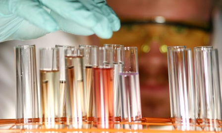
Every analysis of a suspected controlled substance should consist of at least two tests. The first is a presumptive or screening test which indicates if the sample could be a controlled substance. The State Crime Lab's procedures stated in 2008 that presumptive tests are "used to evaluate evidence in determining the possible presence of controlled substances into general categories." (That language has since been removed from lab procedures.) In some cases, crime labs perform only a screening test and then write in the lab report that further testing will be conducted at the request of the District Attorney. Substances other than controlled substances may produce positive results with these tests (false positives). Therefore, confirmatory tests that are substance-specific must be performed in order to positively identify the substance. The State Crime Lab's procedures in 2008 stated that confirmatory tests are "used to conclusively identify the identity of a controlled substance." (This language has since been removed from lab procedures.) The State Crime Lab's procedures divide drug testing techniques into three categories and describe what combinations of tests must be performed. (p. 8) The State Crime Lab's Technical Procedure for Drug Chemistry Analysis provides helpful flow charts that show which test are used to analyze suspected controlled substances.
The State Crime Lab also provides guidelines for proper recording of analytical data (See Sections 5.6). Also see the lab's sampling procedure.
For practical tips on reviewing lab reports, challenging tests, and working with experts, see the papers written by Diane Savage and Dean Loven.
Explanation of Testing Procedures
Detailed descriptions of the tests used to analyze suspected controlled substances are contained in the expandable sections below. Information including links to the State Crime Lab's testing procedures, explanations of how tests are performed, and limitations of the tests are contained in the expandable sections.
- Presumptive Tests
- Color Tests (click to expand)
- Instructional Videos for Chemical Color Tests:
- The Home Scientist – Forensic Presumptive Drug Testing
- Ohio University Forensic Chemistry Lab – Chemical Color Tests
- Concerns for color tests: (1) False positives. Some over the counter cold medication or other substances may test positive for illegal substances using presumptive color tests. Because some drugs behave similarly, a confirmatory test is needed. (2) Color tests may only give an indication of a class of drug present. (3) A color test cannot conclusively identify the presence of a substance.
- Case Law: In State v. Carter (2014), the NC Court of Appeals found the trial court abused its discretion by allowing an officer to testify that a narcotics indicator field test kit indicated the presence of cocaine on items of evidence where the State failed to demonstrate the reliability of the kit.
- To reduce the variability in results due to subjective analysis, good laboratory practices include running a positive control for visual comparison with the evidence sample.
- Typically a lab report from the State Crime Lab will only include the name of the test and the resulting color (or may simply state that the expected color change occurred).
- Link to the State Crime Lab's procedure - Note the quality control check that is required on p. 2.
- Law Enforcement and Corrections Standards and Testing Program - National Institute of Justice standards for color test reagents and kits for the preliminary identification of controlled substances.
- Helpful questions to consider: Was the required quality control check performed? The reagent lasts for only X days - do you know the age of the reagent? How was the resulting color measured - was it compared to a color standard or simply eye-balled? Was the resulting color recorded (photographed)? What precautions were taken to avoid cross-contamination? If performed by a law enforcement officer, what training does he/she have to perform this test?
- Law enforcement officers may use color tests to check for controlled substances while in the field. They typically use a kit that is produced commercially, for example, by NC company Sirchie. An attorney may have to determine which of the tests below is in the kit.
- Marquis Reagent (click to expand)
- What it is: a spot-test to presumptively identify alkaloids as well as other compounds.
- How it works: 1-2 drops of the reagent (a mixture used in chemical analysis) is put on a plate. Approximately 0.1 mg of the unknown powder or tablet is added to the reagent. The color produced is observed and recorded.
- Substances it tests for: MDMA (Ecstasy), opiates (e.g. codeine, heroin), and phenethylamines (e.g. methamphetamine, amphetamine)
- Positive results: opiates – pinkish purple; phenethylamines – dark purple to yellow
- Case law: State v. Powell, 11 N.C. App 365 (1971) a law enforcement officer would need to be qualified as an expert witness in order to testify to result of this test.
- Considerations:The reagent lasts for only 30 days - do you know the age of the reagent?
- Cobalt Thiocyanate Reagent (Scott's Test) (click to expand)
- What it is: widely used to determine the presence of cocaine salt, and is sometimes used to detect heroin and methamphetamine.
- How it works: 2% Cobalt Thiocyanate is dissolved in water and then diluted in 1:1 ratio with glycerin. The solution is then concentrated with hydrochloric acid (HCl). Chloroform is added in the last step. Observation of a blue pigmentation in the chloroform layer is a positive result.
- Substances it tests for: cocaine, heroin, methamphetamine, and PCP
- Positive results: a blue color will be observed in the chloroform layer for the following drugs:
- Cocaine
- Heroin
- Methamphetamine
- PCP
- Considerations: Heat may have a substantial effect on the results. Heat causes the reagent's sensitivity to decrease. May also produce a false positive in the presence of Demerol or other non-controlled substances.
- Duquenois-Levine (click to expand)
- What it is: screening reagent for the presence of marijuana and other cannabinoids
- How it works: If marijuana or cannabinoids are present, a violet-blue color will appear. When chloroform is added, the color will be transferred to the chloroform layer.
- Substances it tests for: cannabis (i.e. marijuana, hashish, hashish oil, THC)
- Positive results: cannabis - violet blue/black
- False Positives: for a list of false positives, see first link below. False positives may occur in the presence of Advil, Nuprin, aspirin, and some brands of coffee.
- Case law: State v. Tate, 300 N.C. 180 (1980). Trial judge suppressed the results from Duquenois-Levine color test because of its propensity to give false positives for coffee and aspirin. NC Supreme Court affirmed.
- Considerations:
- Different procedures are required for wet, fresh, and old plant material.
- If reaction is slow, heat may be added to speed up the reaction.
- For devices or paraphernalia, extra chloroform is added to wash away the marijuana residue. Only a portion of the rinsed chloroform is used for testing.
- Links:
- Ferric Chloride (click to expand)
- What it is: Color test which reacts with GHB, phenols, and enols to produce color changes.
- How it works: Add reagent to well plate and add unknown substance. Observe color change. Can be used for drinks and urine samples.
- Substances it tests for: GHB, phenols, and enols (Some sedatives and anesthetics are phenols or enols.)
- Positive results:
- Phenols - red/brown
- Enols - red/brown
- GHB - red/brown
- Considerations: Acetaminophen is used for Quality Control check. Will produce a violet-purple color.
- Links:
- Koppanyi (click to expand)
- What it is: Color test which reacts with barbiturates to create a color change.
- How it works: Koppanyi paper is created by dissolving cobalt acetate in ethanol. Acetic acid is added to the solution. Paper is then soaked in solution. After paper dries, a small amount of sample is placed on paper. Isopropylamine is dropped above the sample on the paper and tilted to run towards the unknown sample. The color change is observed.
- Substances it tests for: barbiturates
- Positive results:
- Barbiturates - red-violet
- Oxymorphone HCl - violet
- Pseudoephedrine HCl - light green
- Psilocybin - blue
- Theophylline - violet
- Considerations: A known barbiturate is used for quality control purposes. Upon addition of the Isopropylamine reagent, the barbiturate will turn the Koppanyi paper red-violet.
- Potassium Permanganate (click to expand)
- What it is: color test that reacts with double bonds to create a color change.
- How it works: Potassium permanganate and the sample are combined in a culture tube and a color change is observed.
- Substances it tests for: opiates
- Positive results: opiates - brown
- Considerations: A culture tube is recommended instead of a well plate.
- Links:
- Alkenes and Potassium Manganate - scientific explanation of this test
- Ehrlich's, Van Urk's, or p-dimethylaminobenzaledhyde (PDMAB) (click to expand)
- What it is: This test uses a filter paper soaked in reagent. This reagent-soaked paper reacts with indoles (LSD), primary aromatic amines (procaine), and carbamates (organic nitrogen-containing compounds) to produce color intermediates.
- How it works: A small of sample is placed on the PDMAB paper, and a drop of methanol is added. A drop of hydrochloric acid is next dropped on the sample by one of three methods.
- Substances it tests for: LSD, mushrooms (i.e. psilocin, psilocybin), indoles, aromatic amines (procaine), carbamates
- Positive results:
- LSD - purple
- Psilocin - dark purple; psilocybin - red-brown
- Indoles - purple
- Aromatic amines (some antidepressants) - yellow-orange
- Carbamate - yellow
- Considerations: For QC purposes, procaine or LSD can be used. Procaine will produce a yellow-orange color upon addition of hydrochloric acid to the PDMAB paper, while LSD will turn a bright purple.
- Froehde's (click to expand)
- What it is: Molybdic acid (or sodium molybdate) is combined with sulfuric acid by heating and stirring.
- How it works: This reagent will react with many aromatic compounds to produce color intermediates. The aromatic compound will undergo oxidation/reduction/substitution reaction to produce colored intermediates.
- Substances it tests for: opiates
- Positive results:
- Acetaminophen - blue
- Bufotenine - yellow/brown
- Heroin HCl - purple
- MDMA - yellow/dark green to dark blue
- MDA - green to olive to blue
- Morphine - purple
- Considerations: Guaifenesin is used for quality control purposes and will produce a purple color. The reagent expires after 30 days.
- Mecke's (click to expand)
- What it is: Color test that reacts with aromatic compounds to produce colored intermediates.
- How it works: Selenious acid is added to concentrated sulfuric acid while stirring.
- Substances it tests for: aromatic compounds, opiates, amphetamines, heroin
- Positive results:
- Bufotenine - brown to black/purple
- Diphenhydramine HCl - yellow
- Heroin HCl - green/blue
- Hydrocodone bitartrate - dark blue
- Methadone - green/brown
- MDMA - green to dark blue
- MDA - green
- Considerations: The lab's procedure says guaifenesin is used for quality control and produces a green to red color. It is unclear what "green to red" color looks like. The reagent expires after 30 days.
- Other tests include: (click to expand)
- Silver Nitrate for halides. A white precipitate indicates a positive result.
- Zwikker (cupric sulfate, pyridine, chloroform) for barbiturates. A positive result includes a bright purple or bright green color in the organic layer.
- Barium Chloride for sulfates. A white precipitate indicates a positive result.
- Methanol Potassium Hydroxide for cocaine
- Physical Examination (click to expand) - includes techniques (visual examination and microscopic examination) to understand the physical properties and characteristics of substances.
- Visual Examination - for pills, NCSCL's procedure is for analysts to identify a pharmaceutical preparation by physical characteristics and markings. This means the pill is compared to pictures and descriptions in reference materials such as Micromedex, The Physician's Desk Reference, The Logo for Tablets and Capsules or websites. Visual examination is to be used for "preliminary examination only." (p. 7) In State v. Ward the NC Supreme Court held that visual identification of controlled substances is not reliable enough to be admitted in criminal trials, and that a chemical analysis is required. For further discussion see the various posts on the subject on the NC Criminal Law blog. SWGDRUG allows pharmaceutical indicators as a class B identification of pills. This means that 2 other identification methods are necessary in order to positively identify a pill as a controlled substance.
- Microscopic examination - using a microscope to examine the characteristics and properties of a substance too small for the naked eye.
- Polarized light microscopy – contrast-enhancing technique that improves the quality of the image obtained by equipping these microscopes with a polarizer and an analyzer (second polarizer).
- Microcrystalline test – a substance reacts with a reagent forming an insoluble crystal. The shape of the crystal suggests the type of drug. These tests are rapid and do not require the isolation of the drug prior to testing.
- Polarized light microscopy with microcrystalline tests can be used to presumptively identify drugs. The following list indicates various reagents that can be exposed to the drug for analysis using the microcrystalline test with microscopy. Proper reagent preparation and procedures are provided by the NC State Crime Lab Guidelines.
- Heroin and caffeine: mercuric chloride, hydrochloric acid
- Barbiturates: Wagenaar reagent (cupric sulfate), sodium hydroxide, sulfuric acid
- Cocaine and phencyclidine (PCP): gold chloride in acetic acid, hydrochloric acid
- Plant particles from marijuana, hashish: concentrated sodium hydroxide, petroleum ether, chloroform, chloral hydrate
- Amphetamines and methamphetamines: gold chloride in water, gold chloride in phosphoric acid, sodium hydroxide
- Excipients and diluents: hydrochloric acid, methanol, distilled water
- Propoxyphene: acetic acid, gold chloride in acetic acid
- Concerns: (1) Impurities may cause unusual crystal formation. (2) Check reagents with known standard of drug before performing an actual procedure. (3) Proper storage of reagents.
- Macroscopic examination – examining the properties and characteristics of the substance that are observable by the naked eye.
- For descriptions and images of illegal drugs, see the following sources:
- DEA Resource for Parents - provides images of drugs of abuse and information about the effects and legal status of drugs.
- DEA Resource Guide - provides images of drugs of abuse as well as a list of federally scheduled drugs and descriptive information about the major classes of drugs.
- The University of Texas at Arlington – Pictures of Commonly Abused Drugs
- For descriptions and images of illegal drugs, see the following sources:
- For Marijuana - The United Nations Office on Drugs and Crime recommends a combination of color test, thin-layer chromatography and physical (macroscopic and microscopic) examination as a minimum analytical approach for the identification of cannabis products.
- NCSCL Technical Procedure for the Identification of Marijuana
- Polarized Light Microscopy - for viewing suspected hashish/THC samples with the polarized microscope.
- Confirmatory Tests
- Gas Chromatograph/Mass Spectrometer (GC/MS) (click to expand)
- Link to the State Crime Lab's procedure
- Characteristics
- Specific
- Considered the "gold standard" of drug identification
- Most definitive and reliable of the confirmatory tests
- How it works
- GC
- A sample is injected via a port into the gas chromatograph and converted to gas form (vaporized). The vaporized sample is then transported by helium gas through a long coiling column. The column is then subjected to varying temperatures which results in the separation of compounds based on volatility (how easily a substance can be vaporized). Eventually, all molecules will reach the end of the column where they enter the detector. After detection, a computer program will generate a chromatogram, which will show peaks for each of the molecules separated by the GC. The higher the quantity of molecules of the same kind that reach the detector, the higher the peak for that molecule will be. The same is true for the inverse as well.
- The amount of time it takes for a molecule to move through the entire machine and be detected by the detector is called the retention time. Analysts may use these retention times to differentiate between different compounds. However, the retention times alone are not reliable indicators of what compound is present. A specific compound will have a known retention time, but multiple compounds may have the same retention time in the same chromatogram. In order to distinguish and identify which compounds made the peaks, a mass spectrometer is used.
- MS
- A mass spectrometer will determine the exact molecular weight of a molecule or compound. A previously separated sample (flowing directly from GC after detection) or a pure sample (injected) must be ionized for mass spectrometry. The sample, now broken down into molecules, is subjected to an ionization source, a beam of steadily flowing electrons, which blasts the molecules and causes them to break apart into positively charged particles, or ions. Only positively charged ions will continue through the machine. Next, the ions travel through the mass analyzer, or quadrupole filter. The mass analyzer uses an electromagnetic field to separate the ions based on molecular weight. In a routine drug analysis performed by the laboratory, the analyst will already know the molecular weights of the drugs that are frequently detected. It is unnecessary and impractical to know the molecular weight of every compound in a substance. Therefore, to isolate these specific drugs at their known molecular weights, the scientist will manipulate the experimental environment (such as flow rate, type of gas used, temperature, etc.) in previously tested and scientifically accepted ways to produce the range of molecular weights required for analysis. This manipulation will allow only ions between the desired molecular weights to pass through the machine. Ions that flow through the mass analyzer without being filtered will reach the mass detector. The mass detector will count the number of ions with a specific mass. These detections will be translated by a computer program into a mass spectrum.
- Together
- A chromatogram (from the GC) and a spectrum (from the MS) can be used together to determine a molecule's identity. Solely using GC results to identify a compound may lead to inaccurate conclusions. As mentioned earlier, a molecule will have a specific retention time, but a specific retention time does not identify a specific molecule. The analyst will use the spectra to determine the molecular weight of the molecule that generated the peak at a specific retention time. The molecular weight at that retention time will eliminate incorrect identities and narrow down the potential identities to one. The retention time, molecular weight, and patterns of both the chromatogram and spectrum must all match a known standard to be conclusively identified. Differences should not be disregarded unless specifically permitted in the lab procedures.
- Limitations
- A mass spectrometer cannot distinguish between compounds in mixed samples. A sample must be separated prior to MS testing. A gas chromatograph is used to separate the impurities out of a sample in preparation for mass spectrometry. The mass spectrometer will measure the atomic weight of the compounds that were separated by the gas chromatograph.
- MS cannot differentiate between molecules that have the same molecular weight but have chemical structures that are mirror images (also known as enantiomers). This is the case specifically with pseudoephedrine and ephedrine. Subsequent FTIR testing may be necessary to distinguish enantiomers.
- GC/MS will detect the presence of methamphetamine, however, it will not differentiate between methamphetamine stereoisomers. L-Methamphetamine is the active ingredient in the over-the-counter cold medication Vick's VapoInhaler. This ingredient has different effects on the human body and is completely legal. It is commonly labeled as levmetamfetamine to signify that is the legal form of methamphetamine. The other isomer, D-Methamphetamine, is not legal, but will create the exact same peaks on a GC/MS as the L-form does. It is important for the lab to quantitate how much of each stereoisomer are present, which can be done by means of percentages. Some laboratories will allow up to 20% of the D isomer to be present before reporting positive test results for illegal methamphetamine. This blog post explains the concepts of chirality and sterochemistry as they apply to GC/MS testing of suspected methamphetamine.
- If a sample contains a high concentration of pseudoephedrine or ephedrine, it is possible for an "artifact peak" to appear on a GC/MS chromatogram. If not correctly identified as an artifact peak, the sample may be misidentified as methamphetamine. This peak is created by a reaction at the injection point under certain experimental conditions. At higher temperatures (see Hornbeck et al, "Detection of GC/MS Artifact Peak as Methamphetamine," Journal of Analytical Toxicology (1993) which determined that at 300 degrees an artifact peak existed, while at 185 degrees it did not) pseudoephedrine and ephedrine will react with 4-carboxyhexafluorobutyrl, pentafluoropropionyl, heptafluorobutyryl, and a few other substances to create an artifact peak that is similar to that of methamphetamine. This artifact peak can be eliminated by adding sodium periodate to the urine sample before GC extraction.
- Malfunctions in the equipment can occur. The injection point septum is a part of the machine that has the potential to wear. This port only lasts 100-200 injections. The injection port temperature, which is selected by the analyst, may be high or low causing a shortened septum life span, decreased sensitivity, poor separation of liquid material, or decomposition of the sample.
- Considerations
- Is the machine is properly maintained and calibrated? Were the samples prepared properly by the analyst? Are any reagents out-of-date? Were negative controls, positive controls, and blanks properly used? Was the analyst following the protocol properly?
- Did the analyst perform an individualized interpretation or did s/he rely on the computer generated comparison to the library of standards?
- Interpretation of only the chromatogram (chart generated by the gas chromatograph) or a spectrum (chart generated from the mass spectrometer) is not sufficient. Both the chromatogram and spectrum must be interpreted together and a final identification must be supported by both.
- Links
- Webinar on Mass Spectrometry by Forensic Science Initiative, a Continuing Education program from West Virginia University
- Gas Chromatography and High Performance Liquid Chromatography (click to expand)
- Characteristics
- Non-specific
- Used for molecules with high molecular weights.
- Similar to GC, but where the GC is filled with gas, the HPLC column is filled with liquid solvent or solvent mixture.
- The State Crime Laboratory uses this technique for quantitation of methamphetamine in drug analysis cases.
- How it works
- HPLC can be used for qualitative identification: figuring out what is in a sample. HPLC is a separation technique that uses a stationary and mobile phase, a pump, and a detector to separate the compounds in a mixture. The result of the process is a computer generated graph known as a liquid chromatogram.
- First, a small portion of the sample (approximately 5 to 20 µL) is injected into the stationary phase or column. The stationary phase is a small tube, also known as a column that is 3-5 microns in diameter and packed with microscopic particles. Next, the liquid mobile phase is pumped by the exterior pump through the machine. The mobile phase is similar to the gas flowing through a GC. The liquid mobile phase flows through the stationary phase similarly to how water flows through a water filter. The stationary phase separates the molecules in the sample, carrying them down the column at different rates. Eventually, the molecules will travel through the column and reach the detector at the end. The detector will record which molecules are present, and also will record the number of molecules of the exact same chemical makeup are detected. As with a GC, the time it takes the molecule to travel through the column and be measured by the detector is called the molecule's retention time. Similarly, the greater the number of molecules present in the sample, the higher the intensity of the peaks.
- HPLC can also be used for quantitative measurement: figuring out how much of a compound is in a sample. A calibration curve is used to calculate the concentration of the sample.
- Limitations
- The standards used in both GC and HPLC are measured using certain conditions. The analyst chooses the speed of the gas or liquid that travels through the column. They also choose the temperature of the column for each experiment. The temperature of the column, the gas/liquid flow rates, and other factors affect the results. To recreate the experiment and replicate results, the exact conditions chosen by the analyst must be known. The same flow rate and temperature must be used if a second analyst wants to receive the same results on the earlier tested sample. If an analyst wants to compare their results to a known standard sample, they must have performed the experiment under the same conditions that the standard was performed under. As a whole, HPLC results are not as reproducible as GC results.
- This animation shows how these conditions can change the results in an HPLC chromatogram.
- Considerations
- A single HPLC chromatogram cannot be used to calculate the concentration of the compound of interest. A calibration curve must be used to calculate the concentration of the sample. The calibration curve must also be within the accepted ranges allowed by the experiment (QC requirements). Always check that the QC and calibration curve were in range. A calibration curve with a correlation coefficient or R2 value equal to 1.000 is the most desirable. An R2 value of greater than 0.995 is required by the State Crime Laboratory for accurate reporting.
- An HPLC machine cannot test all ranges of concentrations. For a highly concentrated drug, a sample (here referring to a small portion of the overall evidence) will be taken from the whole and diluted to measure the diluted sample's concentration. A concentration for the diluted sample will be calculated using the concentration curve mentioned above. The diluted sample's concentration will be multiplied by the dilution factor to find the calculation of the entire evidentiary sample. The concentrations used in the calibration curve must encompass the concentration of the diluted sample. For example, if the analyst is reporting the concentration of the diluted sample of methamphetamine to be 55 mg/mL, and the standards used to create the calibration curve range from 0.5 mg/mL to 50 mg/mL, then the concentration is out of range and cannot be used. The analyst must either further dilute a second sample to be within the range of the calibration curve or create another calibration curve. The curve's limits may not be extrapolated to determine a sample's concentration. Only the concentrations within the range of the concentration curve are valid.
- Fourier Transform Infrared Spectroscopy (click to expand)
- Link to the State Crime Lab's procedure
- Characteristics
- Highly specific.
- Many substances will create an unequivocal IR spectrum that is easily identifiable by analysts.
- An IR spectrum by itself does not provide an exact chemical structure of a compound, but will provide information about functional groups that are present in the molecule.
- Presence or absence of certain functional groups will guide an analyst as to the possible identity of a compound.
- How it works
- Infrared light is light that is not visible to the human eye. FTIR measures the amount of infrared light that is absorbed by a sample. Molecules (multiple atoms held together by bonds) are constantly moving. The bonds that hold the atoms together will bend, rock, stretch, compress, or twist. These actions collectively are called vibrations. Different functional groups, mentioned above, will absorb infrared light differently and will cause the atoms to vibrate more or less frequently. How quickly the molecule vibrates is called the frequency. More frequent vibrations will create a higher frequency, and fewer vibrations will generate a lower frequency.
- An infrared light beam, or source, is aimed directly at an instrument called an interferometer. The interferometer uses a combination of a beam splitter, a mobile mirror, and a stationary mirror to separate the beam of light into individual wavelengths. First, the beam splitter splits the beam of infrared light at right angles into two smaller beams. One beam will hit the stationary mirror and the other will hit the mobile mirror. The angles of the mirrors allow the two separated beams to reflect, meet again, and reconstitute as one beam. The mirror mechanism separates the light into different wavelengths that are measured at the point of reconstitution. The interferometer records this information and creates a graph called an interferogram. An interferogram records information about every frequency of light from the infrared source. Most importantly, every frequency is measured at once instead of individually as earlier scientific devices required. Once the frequencies of infrared light have been measured by the interferometer, the reconstituted beam continues through the machine to the sample. The beam is applied to the sample where it will either transmit (go through) or reflect (come back towards the source). At this point, specific frequencies of energy will be absorbed by the sample and some will transmit through. The light that does transmit through the source will reach a detector on the other side. Not only does the light that reaches the detector generate information, but the absence of some wavelengths of light, the light absorbed by the sample, also generates information for analysis. The information gathered from the detector is finally sent to a computer where a chart is created specifically for that sample. This computer generated chart is called an IR spectrum.
- Analysts use the IR spectrum and compare the sample's spectrum to a known reference sample. This can be done either by computer comparison, or by an individual analyst manually comparing the two spectra. Each "peak," explained below, is representative of a bond between two atoms. Specific well-known bonds that also have definable physical/chemical characteristics are called functional groups. Common functional groups include alcohols (- OH), ketones (=O), esters (- COOCH3), ethers (-COC-), and carboxylic acids (-COOH), to name a few. Each of these functional groups, if present, will create a distinct peak that typically is easily identifiable by a trained analyst. These peaks will be a specific intensity and be found within a specific wavelength range. When analyzing a spectra, it is important to know that the presence of a peak in a certain place does not necessarily indicate that a specific functional group or bond exists in a sample's chemical structure. However, absence of a peak in the area where a known functional group or bond would present itself does indicate that that functional group or bond does not exist in the sample's chemical structure. Some peaks will be more or less intense due to the bond's nearness to other bonds (the molecule's stereochemistry), the number of the exact same type of bonds present in a molecule (aromatic C-H bonds), and many other more complex reasons. Identification of compounds by IR spectra is an art which takes a lot of practice. Analysts must not only remember the locations of where peaks should be, but must also remember the patterns common to certain types of molecules that could be present in a larger macromolecule. The comparison of the sample's spectra to the known standards is mandatory, either manually or by a computer, and the match must be exact, and forensically, must be supported by another confirmatory test.
- Limitations - limitations of the analysis are based on closely related materials, mixtures, and improper preparation of the sample.
- IR cannot differentiate between isomers of the same drug.
- For an accurate spectrum, the substance must be reasonably pure (generally >90%). Testing of pharmaceuticals using IR may be more successful than the testing of street drugs which are often impure.
- Diamond cell IR, if used, allows smaller amounts of samples to be tested. If diamond cell IR is not used, then the amount of sample required for testing is much higher.
- Considerations
- Peaks from an IR spectrum are read "upside-down." A peak, obviously poorly named, is actually observed at the lowest point on the graph, or the valley of the observed area. IR measures absorption, the opposite of transmission. Absorption and transmission are inversely related: the more a sample absorbs light, less light is actually transmitted through the sample. The more light that transmits through a sample, less light is absorbed by that sample. The y-axis of a spectra measures the percent transmission of a sample. Samples with the highest percent transmission will generate higher (deeper) peaks, and those with the lowest percent transmission will show a lower (shallower) peak or possibly no peak.
- Relative intensities of the bands are important, any mismatch with reference spectrum negates identification. Significant for the identification of the source of an absorption band are intensity (weak, medium or strong), shape (broad or sharp), and position (cm-1) in the spectrum. Many compounds look similar, but an exact match is necessary to confirm the identity of an unknown.
- Links
- Overview of the use of FTIR for analysis of controlled substances.
- FTIR Identification - this blog post by Dr. Fred Whitehurst discusses the limitations of the FTIR for use with forensic samples.
- Organic Chemistry Lecture - by David Van Vranken, Ph.D. of UC Irvine explaining the science behind IR Spectroscopy.
- Video on the science of FTIR
- Video of an FTIR run from start to finish.
- For a fun and rudimentary explanation of light, see Bill Nye The Science Guy's December 24, 1993 Episode on Light and Optics.
- Which Items are Tested?
- When a suspected controlled substance contains many individual packages, crime labs decide which and how many samples to test by following a Sampling Plan.
- The State Crime Lab's Sampling Procedure contains three types of sampling: Administrative, Threshold, and Hypergeometric Sample Selection.
- Administrative Sample Selection is used for pharmaceutical preparations (pills). One pill is chemically analyzed. No inferences are made about unanalyzed material.
- Considerations: the State Crime Lab's procedure does not allow analysts to infer what substances may be present in the untested pills. This means there should not be testimony about the chemical composition of pills that were not chemically analyzed. The analyst may visually inspect the untested pills and compare their appearance to pictures of pills in the Micromedex database. Visual inspection is not sufficiently reliable to identify a substance. State v. Ward, 364 N.C. 133 (2010).
- Threshold Sample Selection is used when it is practicable to test individual analysis of enough units to meet a statutory threshold.
- Hypergeometric Sampling Plan is used for samples with 10 or more packages where threshold sampling is not practicable. The analyst uses statistics to determine how many items to test in order to be able to make an inference about the untested items. The analyst will state that by testing the required number of items she has demonstrated with 95% confidence that the remaining 90% of the material contains the identified controlled substance. Section 4.10.1.1 lists how many samples must be tested for a particular population size.
- Homogeneity: Each package and its contents must be visually inspected for homogeneity of size, weight, color, packaging, markings, labeling, indications of tampering and other characteristics before the samples are subjected to a sampling plan. Section 4.6.2. It may not be possible to detect significant weight variations when visually examining a very small amount of a substance.
- Extrapolation of weight: If the analyzed portion does not meet a weight threshold, additional indiscriminately chosen samples can be weighed to meet the threshold. The lab does not require that those additional samples be tested. Section 4.10.5. The lab allows analysts to extrapolate weight if it is impracticable to obtain individual weights. Section 4.10.5.1.1.
- Previously used method: From 1996-2010, the crime lab and many other forensic labs determined the number of samples to be tested by using the √n+1 formula where n is the total number of samples. The analyst would make an inference about the chemical composition of the remaining samples after testing √n+1 samples. There was no scientific basis for using this method to determine how many samples to test. For additional information on this method, see this paper by Fred Whitehurst, JD, Ph.D.
- Residue amounts will not be tested by the State Crime Lab unless accompanied by a written request from a prosecuting attorney. If the case consists of items that are all residue amounts, analysis will be performed on items until any controlled substance is identified. See Section 4.5.2.1.
Articles and Resources
- Recent drug analysis news articles - this page contains links to recent press coverage of local and national cases involving drug analysis and is updated regularly.
- NC applies strict pleading standards in cases involving drug names. If the substance listed in the indictment is not listed in the drug schedules or is misspelled, take a look at this blog post and the cases referenced and consider whether the indictment is defective.
- Weighing Marijuana Reference - this document provides the relevant statutes and summarizes the case law on the issue of how marijuana should be weighed. It addresses issues such as whether water weight and mature stalks should be included. Links to the State Crime Lab's relevant procedures are provided, as well as contact information of experts who are available to weigh suspected marijuana.
- False Positives Equal False Justice - a California Attorneys for Criminal Justice (CACJ) report by John Kelly. The report is largely based on the research of Dr. Frederic Whitehurst who tested field drug test kits and exposed and documented that they render false positives with legal substances. The report focuses on the Duquenois-Levine and KN Reagent tests used to test for marijuana.
- Forensic Crime Labs: Scrutinizing Results, Audits & Accreditation by Frederic Whitehurst, The Champion Magazine, April 2004 - This article lists each document that you should request and receive in discovery, describes its relevance and suggests questions that attorneys should ask about each item. The first section of the article also gives a history of lab scandals across the country. Available to NACDL members.
- Drug Identification Tool - this website contains a library of GCMS data for compounds and is organized by molecular weight, base peak and second base peak. This library may be useful for limited purposes.
- PubChem - this National Institute of Health searchable database allows users to search for a compound and learn more about its chemical structure, uses, properties, toxicity, and other information.
- Pillbox - this US National Library of Medicine website allows users to learn the possible identity of pills based on their appearance, color, shape, and markings.